A major component of red bone marrow, located within both long bones (in the young individual) and spongy bone, is the population of haemopoietic/hemopoietic cells. Haemopoiesis/Haematopoiesis refers to the formation of red (erythropoiesis) and white (granulopoiesis/leukopoiesis) blood cells and also the formation of platelets (thrombopoiesis). The formation of lymphocytes (lymphopoiesis) also occurs in the bone marrow and then lymphocytes proliferate and further differentiate in the lymphatic tissues and lymphatic organs but this lineage is not yet described in this resource.
Learning outcomes
After viewing the histological images and interactive text in this module you should
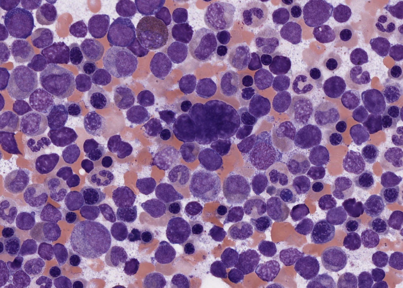
- learn how to examine the cells in red bone marrow smears.
- understand what developing progranulocytes, myeloblasts and lymphocytes look like.
- identify the following stages in the formation of erythrocytes, neutrophils, and eosinophils and understand why the cells change in appearance as they mature:
proerythroblast.
erythroblast – distinguish early (basophilic) from late (polychromatophilic) stages.
normoblast.
neutrophilic metamyelocyte.
neutrophilic leukocyte (mature neutrophil).
eosinophilic myelocyte.
eosinophilic leukocyte.
- observe megakaryocytes (and maybe a megakaryoblast).
- observe that cells of the developing erythrocyte and leukocyte lineages are clustered in bone marrow.
- observe fat cells and macrophages in sections of red bone marrow.