Bone is the ideal tissue to form our skeleton to provide support, movement and protection of our most vital organs. This is because the matrix of bone is very specialized – it is minieralized which make it a very hard tissue.
Learning outcomes
After viewing the histological images and interactive text in this module you should
- recognize bone in a ground section and a decalcified section and visually distinguish adult from woven bone and spongy from compact bone.
- identify in these sections (particularly in the decalcified section):
Osteocytes, lacinae, osteoblasts, osteoclasts.
Bone matrix, osteoid, canaliculi.
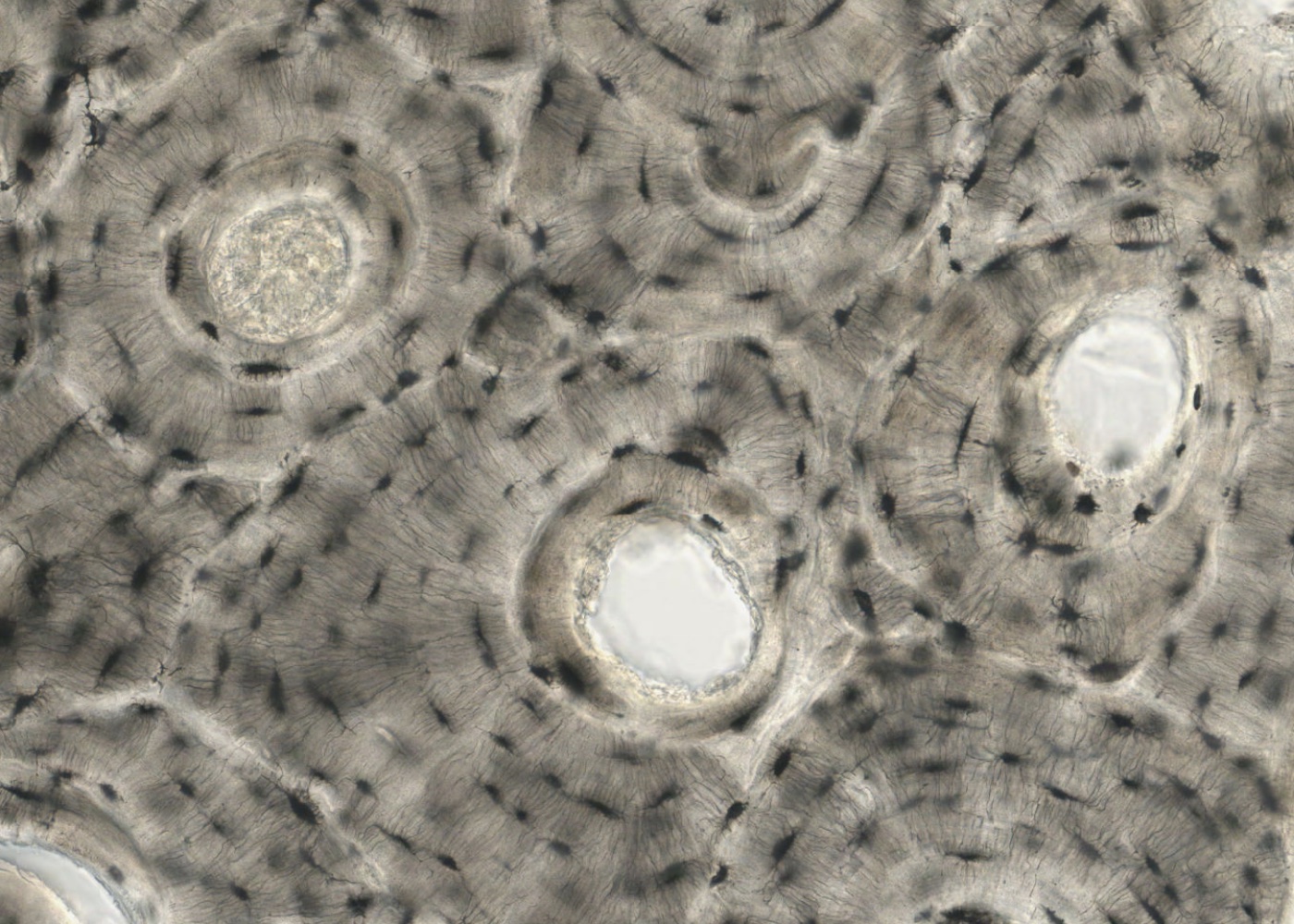
Haversian system (osteon) and Haversian canal.
Circumferential, concentric and interstitial lamellae.
Trabeculum (spicule) of bone.
Periosteum and endosteum.
Epiphysis (epiphyseal plate composed of epiphyseal cartilage) and diaphysis.
- Observe stages of intramembranous and endochondral bone formation and recognize
primary and secondary ossification centres.
reserve cartilage, zones of proliferation, hypertrophy (cell enlargement), calcification. chondrocyte death, resorption and ossification.
spicules of mixed calcified cartilage and bone.
- Understand the histology of bone remodeling.
- Observe the structure of joints:
diarthrosis (articular cartilage, synovial space and synovial organ) and synarthrosis.