Cartilage contains over 95% extracellular matrix in which cartilage cells (chondocytes) are embedded. This matrix is specialised in the different types of cartilage (hyaline cartilage, elastic cartilage and fibrocartilage) to effect their different functional roles.
Learning outcomes
After viewing the histological images and interactive text in this module you should
- Observe the characteristic features of cartilage, including the perichondrium.
- Recognize:
hyaline cartilage and articular cartilage.
elastic cartilage.
fibrocartilage.
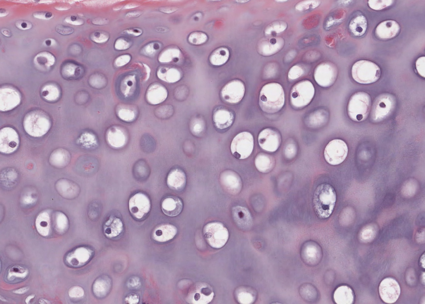
chondocytes in lacunae.
chondrocyte territorial matrix.
interterritorial matrix.
cell nest (isogenous group).
perichondrium – fibrous and chondrogenic layer.
- Understand the functional roles of each type of cartilage and where each type is located in the human body.