Histology Atlas Curriculum

Connective tissue

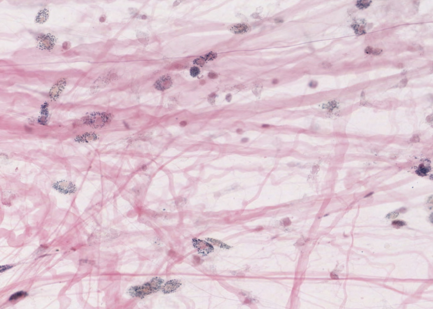
Connective tissue is widespread throughout the body and consists of cells, fibers and matrix. The types of connective tissues depends on which of these three components dominate (functionally).

Learning outcomes

You should be able to identify the following in tissue sections and appreciate their functional significances:

  • Loose connective tissue.
  • Collagen fibers/fibres.
  • Dense, irregular connective tissue.
  • Dense, regular connective tissue.
  • Reticular connective tissue.
  • Reticular fibers/fibres.
  • Elastic fibers/fibres (when stained).
  • Elastic lamina (when stained).
  • Adipose tissue (yellow fat).
  • Brown fat.
  • Stroma.
  • Fibroblasts.
  • Macrophages when vitally stained.
  • Mast cells.
  • Adipocytes.

Learning Modules

Connective tissue cells

Connective tissue “spread”

Connective tissue fibres

Mesenchyme

Adipose tissue

Lamina propria

Loose (areolar) connective tissue

Reticular connective tissue

Dense regular connective tissue e.g. cornea

Dense regular connective tissue e.g. tendon

Dense irregular connective tissue e.g. dermis

Dense irregular connective tissue e.g. artery wall

Interactive Histology Atlas + Descriptive Videos and Quizzes Special Offer

Sign up for 12 months access to the Interactive Histology Atlas (including Virtual Microscopy) for only $19.99 USD and get a Subscription to the Histology Videos for FREE.