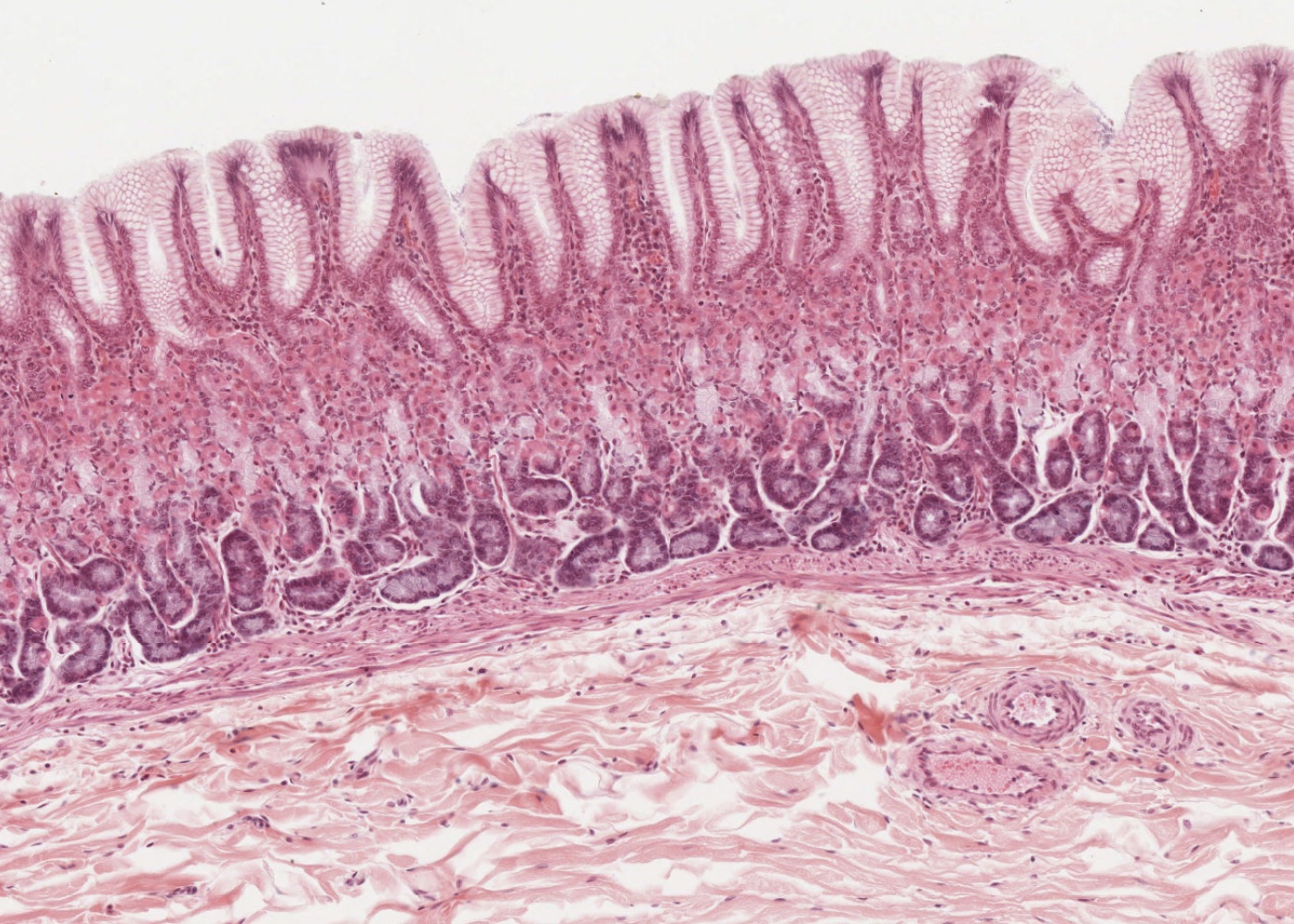
The digestive system includes the alimentary canal, organs within the oral cavity as well as the pancreas, liver and gallbladder. This module covers the principal histological features of the alimentary canal.
Learning outcomes
When you have completed viewing the images and the histological sections available of the organs of the alimentary canal you should
- know the four characteristic layers of the Gastro-Intestinal tract (GI) throughout its length.
- understand how the mucosa, submucosa, muscularis externa of each organ within the GI tract is specialized.
- know the names/histological features of these specializations – particularly the epithelial and secretory cells within the mucosae.
- know what the Diffuse Neuroendocrine System (DNES), Gut Associated Lymphatic Tissue (GALT), and enteric nervous system are.
- describe the main components of the GALT.
- understand the difference between a mucosal and a submucosal structure.
- understand the difference between the mucosal cells of the gastric pits and goblet cells.
- know where the crypts of Lieberkühn are located and what their main functions are.

