Histology Atlas Curriculum

Epithelium

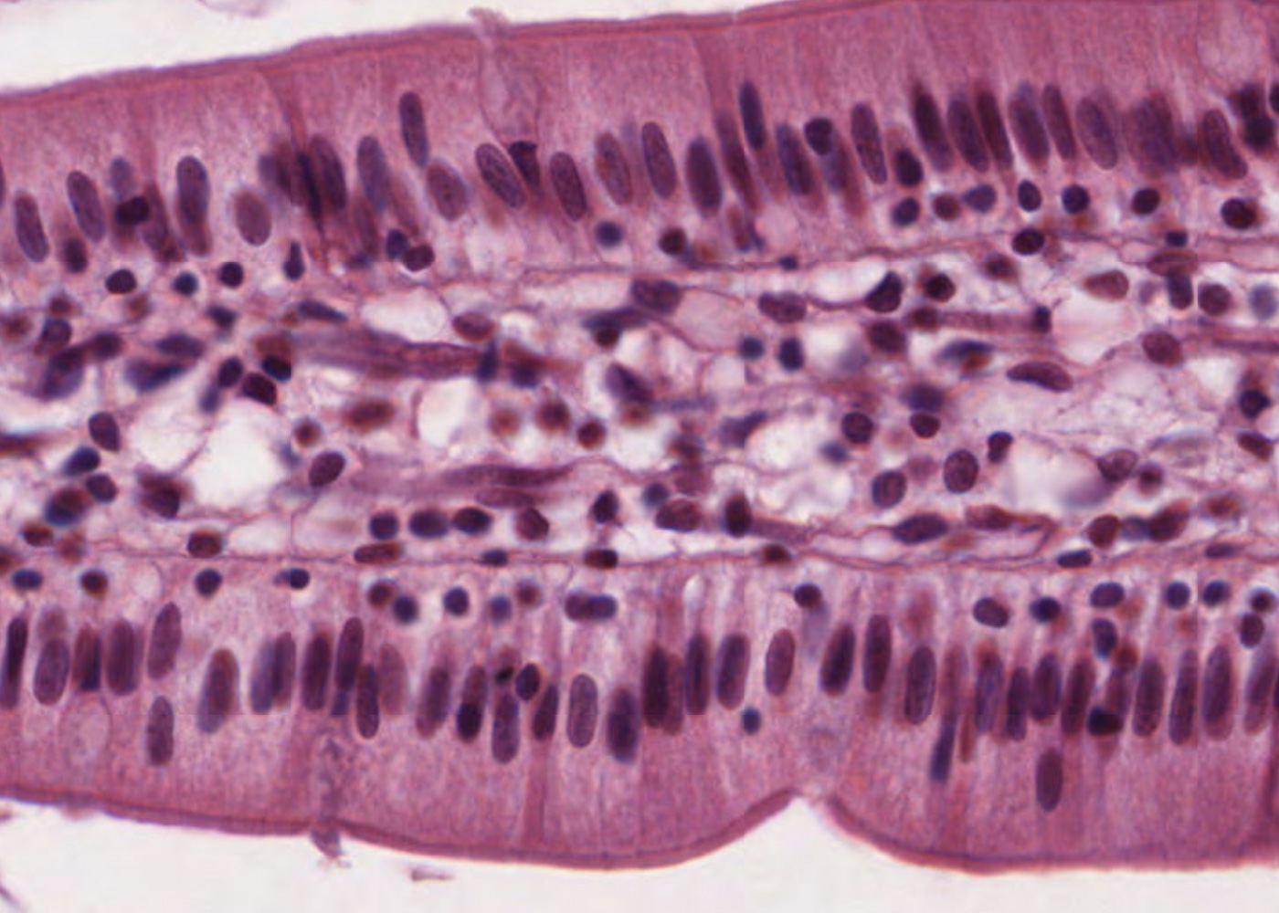
Epithelium is one of the four basic tissues of the body. The other three being muscle tissue, connective tissue and nerve tissue. Epithelium is located in most organs of the body and an understanding of the different types of epithelia, and their respective functions, helps you understand functions of organs and organ systems.

Learning outcomes

After viewing the histological images and interactive text in this module you should

  • identify and distinguish between all the different classifications of epithelium.
  • identify different types of epithelia in various tissues.
  • know the characteristics of epithelia.
  • identify and describe the various types of cell adhesions and intercellular contacts.
  • understand the difference between a basal lamina and a basement membrane and be able to recognize each.
  • distinguish between microvilli, stereocilia and cilia and know their specific functions.

Learning Modules

Intra-qpithelial cells

Myoepithelium

Endothelium

Neuroepithelium

Mesothelium

Basement membrane

The junctional complex

Surface specializations

Transitional epithelium

Stratified columnar epithelium (penile urethra)

Stratified columnar epithelium (eyelid)

Stratified cuboidal epithelium (exocrine gland duct)

Stratified cuboidal epithelium (eyelid)

Parakeratinised epithelium

Stratified squamous (keratinised; thick) epithelium

Stratified squamous (keratinised; thin) epithelium

Stratified squamous (non-keratinised; thick) epithelium

Stratified squamous (non-keratinised; thick) epithelium

Pseudostratified columnar (with stereocilia)

Pseudostratified columnar (ciliated) epithelium

Simple columnar (ciliated) epithelium

Simple columnar

Simple cuboidal

Simple squamous

Interactive Histology Atlas + Descriptive Videos and Quizzes Special Offer

Sign up for 12 months access to the Interactive Histology Atlas (including Virtual Microscopy) for only $19.99 USD and get a Subscription to the Histology Videos for FREE.