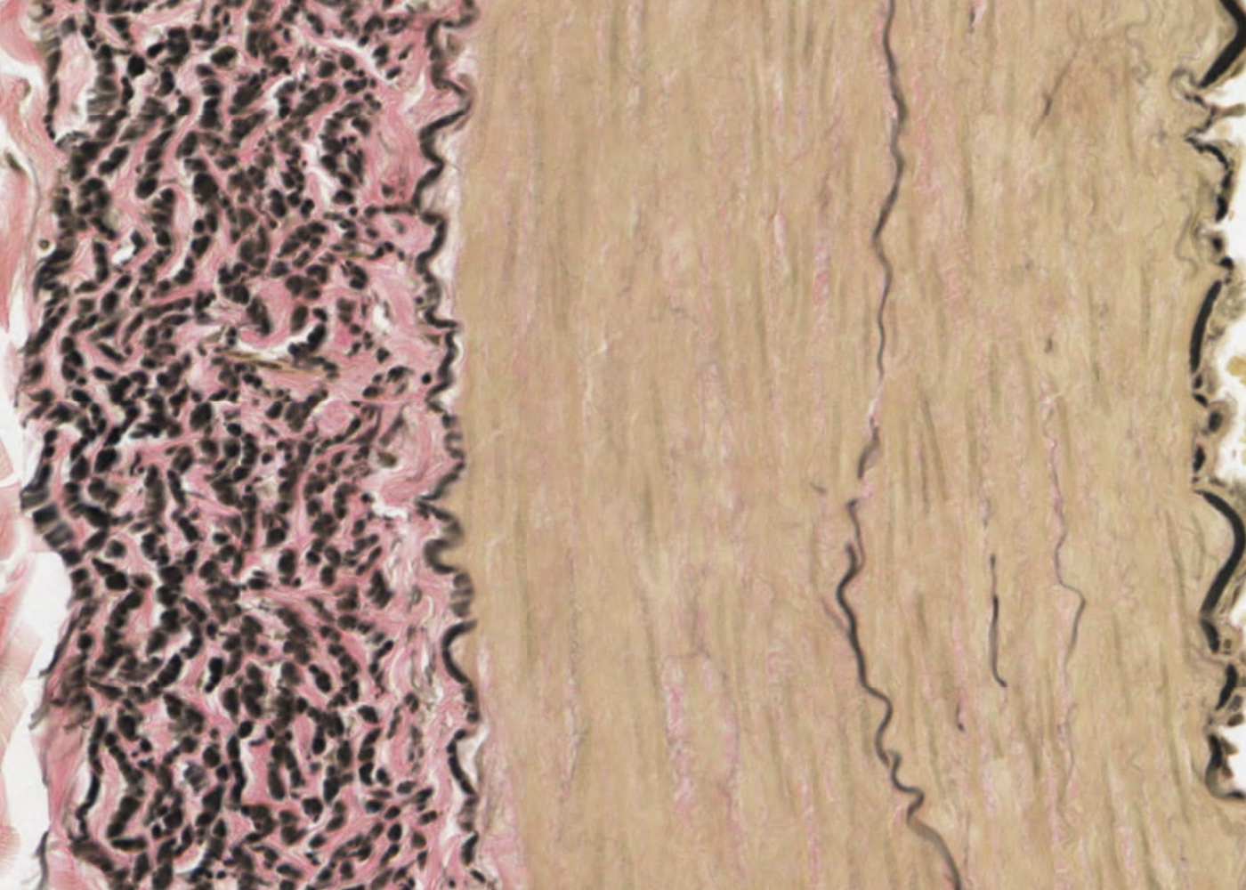
Hematoxylin and eosin are the most commonly used dyes in histology. Eosin is an acidic dye and hematoxylin is a basic dye
Acid dyes react with cationic groups in cells and tissues (particularly with the ionized amino groups of proteins). Basic dyes react with anionic components of cells and tissues (i.e. those carrying a net negative charge)
Anionic components include:
- phosphate groups of nucleic acids
- sulphate groups of glycoaminoglycans
- carboxyl groups of proteins
The ability of these anionic groups to react with a basic dye is called basophilia. Tissue components that stain with hematoxylin also exhibit basophilia.
The reaction of the anionic groups varies with pH. So, staining with basic dyes at a specific pH can be used to focus on specific anionic groups that may be found predominantly on certain macromolecules.
The reaction of cationic groups with an acid dye is called acidophilia. The reactions are not as specific or precise as reactions with basic dyes so often a number of acid dyes are used in combinations. Usually hematoxylin is used first to stain the nucleus then acid dyes are used to stain the cytoplasm and extracellular fibres.
A limited number of substances within cells and the extracellular matrix display basophilia:
- Heterochromatin and nucleoli (ionized phosphate groups in nucleic acids)
- Ergastoplasm and ribosomes (ionized phosphate groups in ribosomal-RNA)
- Extracellular material e.g. matrix of cartilage (ionized sulphate groups in nucleic acids)
Staining with acid dyes is less specific but more substances within cells exhibit acidophilia:
- Cytoplasmic filaments (e.g. contractile filaments in muscle cells)
- Intercellular membranous components (and unspecialised cytoplasm)
- Extracellular fibres (ionized amino acid groups)
Learning outcomes
After viewing the histological images and interactive text in this module you should
understand what specific cell components stain with hematoxylin and eosin.
know the meaning of basophilia and acidophilia.
appreciate various other stains (examples are provided as items in this topic) can be used to distinguish cell and tissue components.

