Nerve tissue controls many of the activities of most tissues and organs in the body. This first module deals with the “peripheral nervous system” and then it is followed by the next topic dealing with the “central nervous system” i.e. the spinal cord and the brain.
Learning outcomes
After viewing the histological images and interactive text in this module you should be able to recognize the following structures:
- Peripheral Nerve
myelinated axon.
nonmyelinated axon.
Schwann cells.
node of Ranvier.
epineurium.
perineurial sheath.
endoneurium.
endothelial cells & fibroblasts.
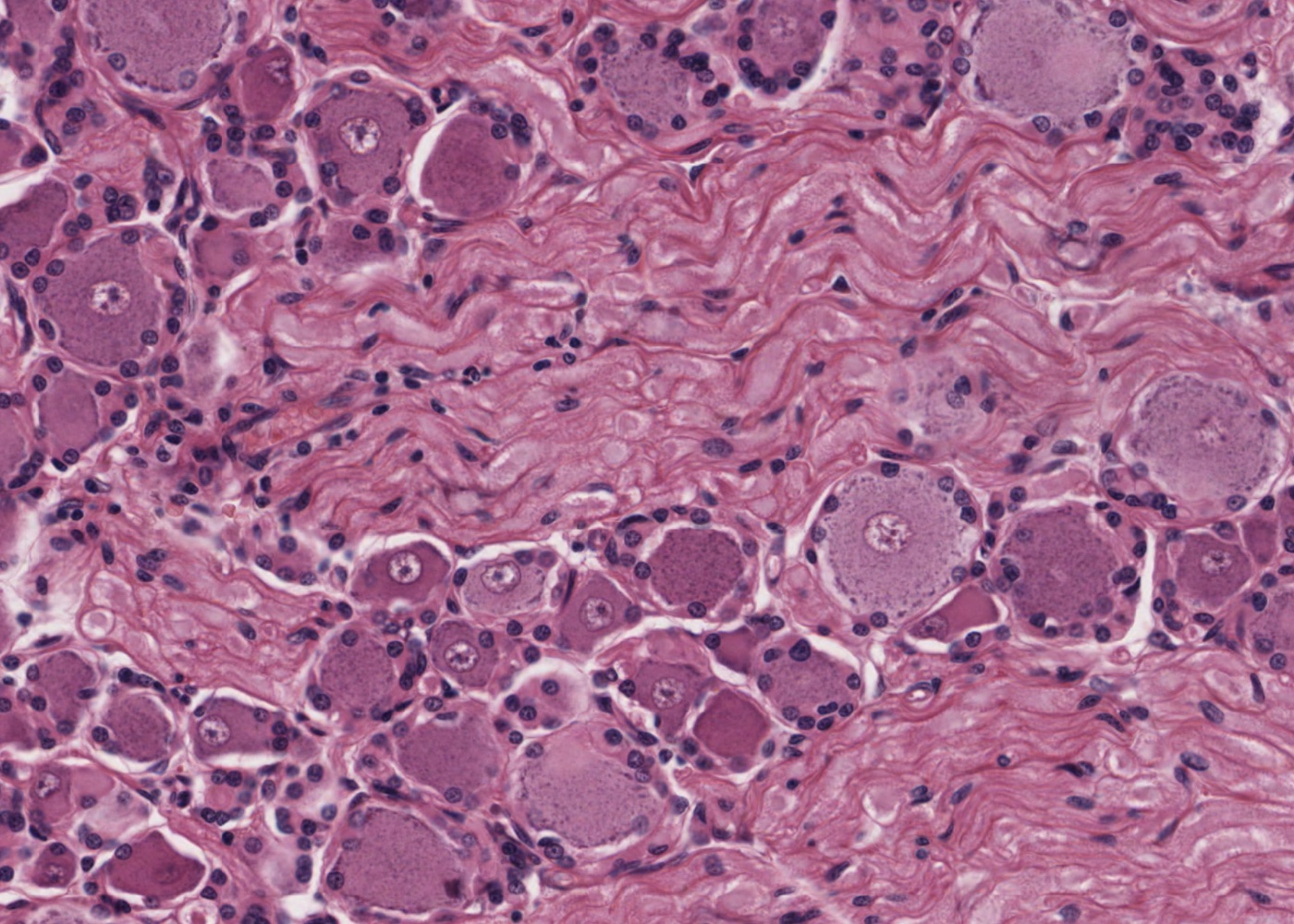
- Sensory ganglion
ganglion cell.
satellite cells.
- Autonomic ganglion
sympathetic ganglion.
multipolar neuron.
postganglionic sympathetic neuron.
myenteric ganglion.
- Distinguish myelinated and unmyelinated axons in peripheral nerves with the following stains:
Masson stain.
Bodian stain.
H&E.
- Recognize small branches of nerves running in connective tissue.