The spinal cord (housed within the spinal canal of the vertebral column) and the brain (protected within the neurocranium) make up the “central nervous system”.
Learning outcomes
After viewing the histological images and interactive text in this module you should be able to recognize the following structures:
- Neuron
cell body.
Nissl substance.
dendrite.
nucleus.
nucleolus.
- Nervous tissue
white matter.
gray matter.
neuropil.
astrocyte.
oligodendrocyte.
ependymal cell.
microglial cell.
endothelial cell.
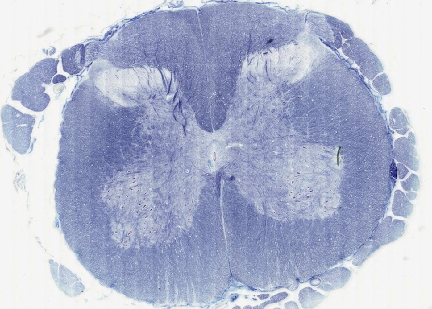
- Spinal cord
dorsal horn.
ventral horn.
lateral horn.
motor neuron.
interneuron.
central canal and ependymal lining.
nerve rootlets.
- Cerebellum
cortex.
molecular layer.
Purkinje cells.
granule cell layer.
granule cells.
- Cerebrum
pyramidal cells.
astrocytes.
- Meninges
dura.
subdural space.
arachnoid.
subarachnoid space.
pia.
- Ventricles
ependymal cells.
choroid plexus.