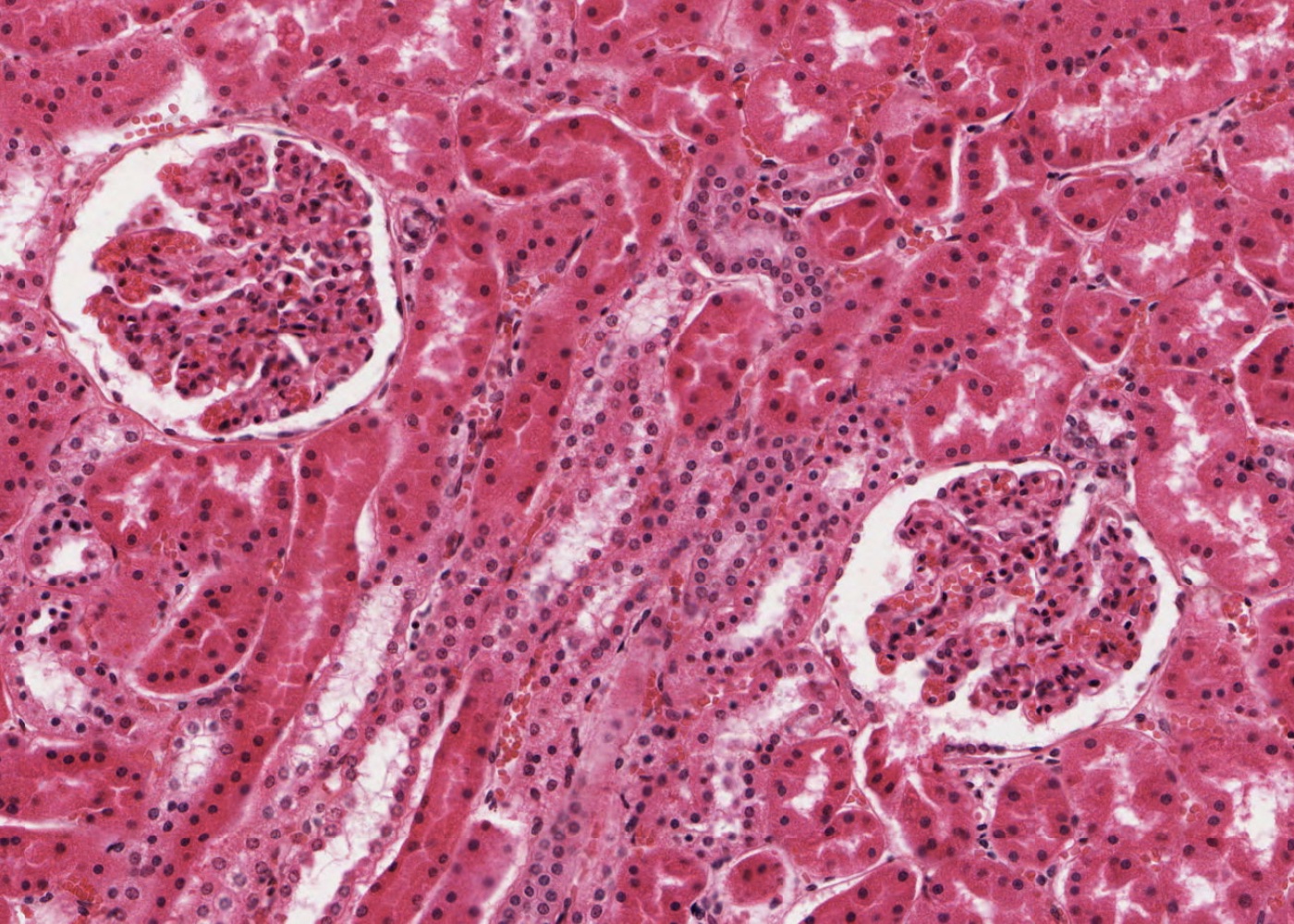
The urinary system consists of the paired kidneys, paired ureters, urinary bladder and the urethra. This system has numerous specific functional roles centred on conserving essential components of the blood and extracellular/body fluids – and eliminating wastes.
Learning outcomes
After studying this module, you should be able to
- recognize the following gross structures of the kidney:
- pyramid.
- lobe.
- major and minor calyx.
- distinguish the medulla from the cortex.
- renal pelvis and ureter.
- renal papilla.
- major arteries and veins supplying and draining the kidney.
- recognize and define a lobule.
- define and describe a uriniferous tubule, a nephron, and a renal corpuscle.
- know the structure and function of the glomerulus and the filtration apparatus.
- identify and understand the general functions of the proximal convoluted tubule, the loop of Henle, the distal convoluted tubule and the collecting ducts.
- recognise a macula densa and understand the function of the juxtaglomerular apparatus.
- understand the functioning of the renin-angiotensin-aldosterone-ADH system.
- understand the blood supply to the kidney and to the nephron.
- recognise the vasa recta and understand their origin.
- differentiate between the cortical and juxtamedullary nephrons and understand the special function of the latter.
- know the general functions of the ureter, bladder and the male and female urethra.

