The interactive histology atlas contains over 2200 high resolution images of all human cells, tissues, and organs. With appropriate technology embedded in this resource to encourage interactive learning, the user’s participation is required when using all the functionalities of this atlas.
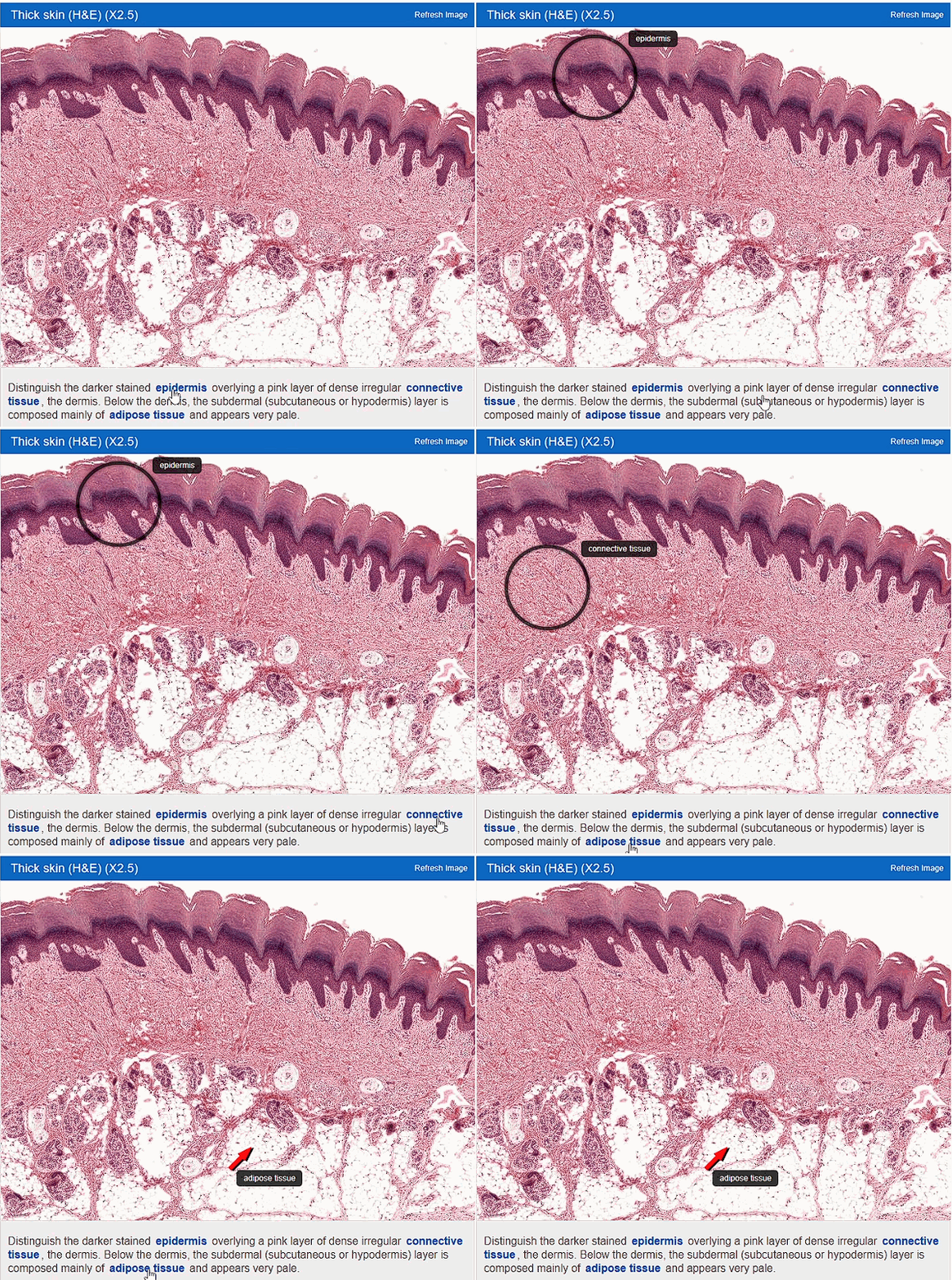
Each image is accompanied by concise descriptive text with named key histological structures hyperlinked. Clicking the mouse on the hyperlink labels the specific histological structure as indicated in the “Interactive Learning Example” in the frame below.
Users can complete laboratory practical exercises by viewing histological sections using the virtual microscopy facility linked to each histological image.

